What are X-Ray and CT Scan Technology?
 By Anolytics | 29 August, 2022 in AI in Healthcare | 3 mins read

An exciting potential to enhance the availability, latency, accuracy, and consistency of X-ray ultrasound or CT scan image interpretation is presented by implementing machine learning (ML) for medical imaging applications. Many algorithms have previously been created to identify specific diseases.
The usefulness of these algorithms in a general clinical situation, where various disorders might manifest, may be constrained by the fact that they were developed to identify a particular illness.
According to the National Practitioner Data Study, in 2011, the use of AI based x-ray and CT scan technology more than doubled, thanks to x-ray and CT scan annotation. The rise of health care technology has also led to an increase in the usage of clinical technologists. This means that more physicians are using x-ray and CT scan technology in their practices.
The artificial inntelligence in medical community is also experiencing a period of growth as more doctors begin to take advantage of x-ray and CT scan annotation technology as part of their standard care delivery strategy. There are many benefits to utilizing these technologies in your practice, including reducing hospitalization, Inefficient workflow, Lower costs, and Efficient no waiting lists.
Let’s explore how you can fit them into your practice settings using the simple steps below.
What are x-ray and CT scan technology?
X-ray and CT scan technologies are based on the same principles as MRIs and PET scans. These technologies use a camera, an accelerometer, and a camera that measures movement, such as walking or running. The camera images are sent to a computer for analysis, and data is collected about the patient’s body composition, energy flow, and other factors.
CT scanning was first developed as a diagnostic tool. Since then, it has become one of the most important diagnostic tools in the medical setting. The medical community has been using CT technology for over 40 years. It is becoming more widespread, with more physicians now using it in their daily practices.
Annotation of a medical image
Medical picture annotation is the foundation of all machine learning research in the healthcare industry. A tremendous amount of such data is required for AI systems to assess and anticipate outcomes confidently, and precisely labeled ultrasound data annotation pictures are necessary to train models exactly. It would be challenging to construct cutting-edge algorithms that might one day be able to detect and anticipate a wide range of illnesses without the focused effort of the hundreds of people working to conduct the tedious, repetitive activities of data annotation.
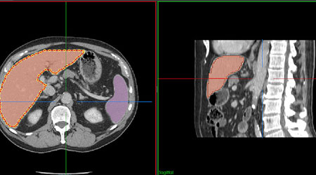
Annotation is a medical imaging technique to highlight areas of interest (often using boxes, circles, or arrows). The word annotation refers to adding metadata to a picture in the related area of digital imaging so that a computer model may be trained to detect particular characteristics. A medical picture annotator often carries out one of two types of annotation.
Segmentation, the first type, involves categorizing individual pixels. Classifying a whole image inside a dataset is the second type. The standard Digital Imaging and Communications in Medicine (DICOM) format is used to edit and encode images. NIfTI is another popular format that creates a 3D picture (as opposed to the single slices format of DICOM). The reader can also change this format, depending on their preferences.
Computerized automated and semiautomatic algorithms are a viable choice in the clinical area due to the ever-increasing volume of patient data collected in hospitals and the requirement to support a patient diagnosed with this data. To train and employ machine learning techniques to simulate a human annotator, such systems for diagnosis assistance must first have manually annotated datasets.
For radiological imaging, the 3D volumes of the patients are manually segmented to separate the different structures in the pictures and enable the processing of the tissue of the systems independently. On the other hand, manual segmentation necessitates arduous and time-consuming work from radiologists.
Conclusion
VR and MR technology have been used in virtual and Augmented Reality (AR) immersive environments for years. These technologies enable real-time 3D mapping and the delivery of software-defined signals.
This approach allows for low-cost, high-power devices that are small and inexpensive to be used in both virtual and augmented reality applications. It’s important to note that these technologies don’t require the presence of humans for practical use.
please contact our expert.
Talk to an Expert →
You might be interested

- AI in Healthcare 27 Mar, 2020
How Big Data & AI is used in Healthcare System to Combat Coronavirus Outbreak?
Coronavirus or scientific name COVID-19, is one the most deadly virus of the century infected more than half of the mill
Read More →
- AI in Healthcare 16 May, 2020
Best Diagnostic Imaging Techniques for Using AI in Medical Diagnosis
Artificial intelligence (AI) and machine learning have enough potential to make various tasks in the healthcare industry
Read More →
- AI in Healthcare 11 Dec, 2020
Best Uses of Artificial Intelligence in Dentistry
Artificial intelligence (AI) is a branch of computer science that describes the research and development of computer-gen
Read More →